If you have been living with knee pain that nobody seems to be able to explain properly, you are not alone. Many patients with plica syndrome spend months seeing different healthcare professionals, having normal X-rays and inconclusive MRI scans, and being told their knee looks fine when it clearly does not feel fine. Plica syndrome is a genuine, specific, and very treatable condition, but because its symptoms closely mimic several other knee problems, it is routinely missed or misattributed to something else entirely.
Plica synovialis is the medical term for a fold of tissue in the lining of the knee joint. When this fold becomes irritated, thickened, and inflamed, it causes a distinctive pattern of anterior knee pain that can feel remarkably similar to a meniscal tear, patellofemoral pain, or general knee inflammation. This is precisely why synovial plica syndrome gets missed so often. The symptoms are real and significant, but they point to a structure that many clinicians do not routinely assess and that standard imaging frequently fails to identify.
The good news is that once plica syndrome of the knee is correctly identified, most patients respond very well to treatment, often without needing surgery at all.
Why Plica Syndrome Gets Missed and What It Actually Is
Plica syndrome gets missed so frequently because it does not behave the way most knee conditions do. It does not always show up clearly on MRI, it does not cause the specific pattern of joint line tenderness that a meniscal tear produces, and it is not something many patients or even general practitioners have heard of. The result is that patients are often told their knee is structurally normal and sent away without a clear answer, when in fact the problem lies in a small fold of synovial tissue that has become chronically irritated.
To understand what plica syndrome is, it helps to know a little about the synovial membrane of the knee. This is the thin, fluid-producing lining that surrounds and lubricates the entire joint. During fetal development, this lining forms temporary folds called plica that divide the developing knee into separate compartments. In most people, these folds shrink and almost disappear before birth. However, around 95 percent of people who undergo knee arthroscopy are found to have some remnant of these folds, most of which cause no symptoms whatsoever. In a smaller proportion of people, one of these folds remains thick enough that it can become irritated, inflamed, and problematic.
What Is a Medial Plica in the Knee
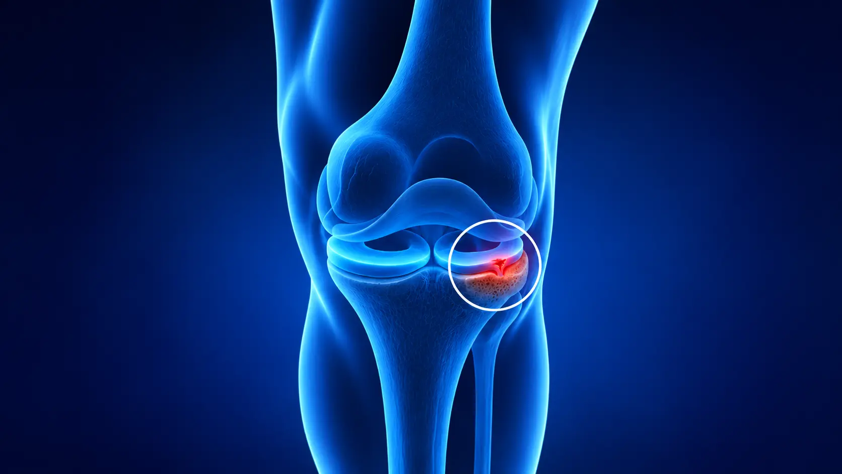
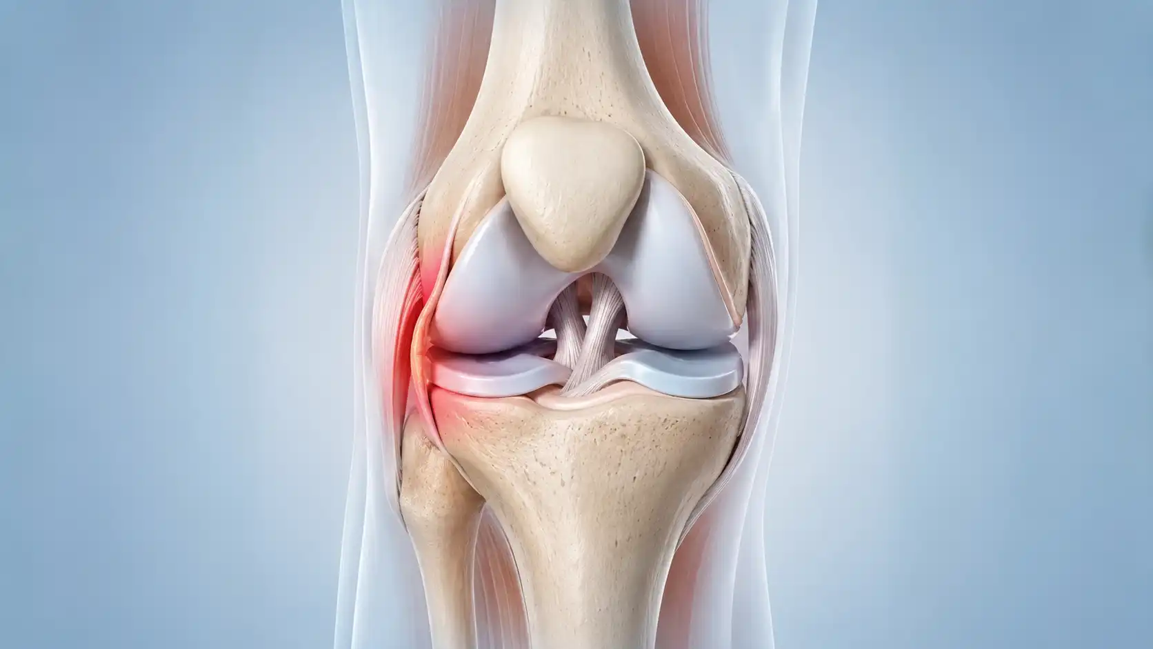
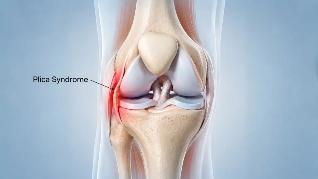
Of the four plica types in the knee, the medial plica is by far the most commonly symptomatic. The medial plica is a fold of synovial tissue that runs along the inner side of the knee joint, between the kneecap and the thigh bone. What is medial plica in practical terms? It is a band of tissue that in most people glides painlessly across the joint surfaces during bending and straightening. When it becomes thickened through overuse or injury, it starts to snap across the inner surface of the thigh bone during movement, causing the characteristic catching, clicking, and inner knee pain that patients describe.
The Four Types of Knee Plica and Which Cause Problems
The knee has four main plica types. The medial plica, as described above, is the most commonly affected. The suprapatellar plica syndrome presentation involves a fold at the top of the kneecap that occasionally causes quadriceps tendon-related symptoms and can occasionally restrict fluid flow within the joint. The infrapatellar plica sits below the kneecap and is less commonly symptomatic. Lateral plica syndrome, involving a fold on the outer side of the knee, is the rarest of the four. Understanding which type is involved helps guide both diagnosis and treatment, though the medial plica accounts for the vast majority of cases we assess and treat.
Plica Syndrome Symptoms and Where the Pain Usually Sits
Plica syndrome pain location is one of the most useful clues in identifying this condition. Unlike a meniscal tear, which tends to cause pain deep within the joint or towards the back of the knee, plica knee pain sits specifically over the inner plica, along the inner edge of the kneecap or just below it. Many patients describe being able to press directly on a tender spot along the inner knee and reproduce their pain immediately, which is a hallmark of medial patella plica syndrome.
The symptoms of plica syndrome develop either gradually through overuse or more suddenly after a specific injury. Common plica syndrome symptoms include the following.
- A dull, aching pain at the front or inner side of the knee that worsens with activity
- Knee clicking or snapping during bending and straightening, often felt rather than just heard
- A catching or locking sensation when getting up after sitting for a long time
- Knee pain when climbing stairs, squatting, or getting in and out of a car
- Inner knee pain that is worse after running, cycling, or repetitive bending activities
- Knee instability from plica syndrome, where the joint feels unreliable on slopes or uneven ground
- Knee pain worse at night due to the build-up of inflammation after a day of activity
- Swelling around the inner kneecap that can sometimes be felt when pressing on the area
The pattern of symptoms that is most distinctive is pain that builds during repetitive activity, eases briefly with rest, but then returns quickly when activity resumes. Knee pain after running is a very common presentation, particularly in patients who have recently increased their training load or returned to exercise after a break.
What Causes Plica Syndrome to Develop
The most important thing to understand about plica syndrome causes is that the plica itself is a normal structure. Does everyone have plica in the knee? Yes, the vast majority of people do. The problem is not the presence of the plica but rather what happens to it when the knee is repeatedly stressed or directly injured.
Overuse is the most common trigger. Plica syndrome causes from overuse occur when repetitive bending and straightening of the knee, during running, cycling, using a stair machine, or performing squats, gradually irritates the plica. Each time the knee bends, the plica snaps across the surface of the thigh bone. In a normal thin plica this causes no problem at all. But with repeated stress, the plica becomes inflamed, thickens, and loses its flexibility. The more it thickens, the more it catches during movement, creating a cycle of irritation that worsens without intervention.
Direct trauma is the second main cause. A blow to the inner side of the knee, for example from a fall onto a hard surface, a sports collision, or hitting the knee on a dashboard in a road traffic accident, can trigger an acute inflammatory response in the plica that then becomes chronic if not properly managed.
Why Plica Syndrome Is So Common in Athletes
Plica syndrome from exercise is particularly common in runners, cyclists, and those who train regularly on steps or inclines. The repetitive flexion and extension pattern of these activities places continuous loading on the medial plica with every stride or revolution. Running plica syndrome tends to develop insidiously, often starting as a mild ache that athletes push through before it becomes persistent enough to seek help. Cycling plica syndrome follows a similar pattern, particularly when saddle height is suboptimal and forces the knee to work through a restricted range with each pedal stroke.
Plica Syndrome Test and How We Diagnose It Accurately
Plica syndrome diagnosis relies more on a careful clinical assessment than on imaging alone, which is one of the key reasons it is so often misdiagnosed when patients are seen only briefly or when a scan result is allowed to override clinical judgement.
The medial plica test is a straightforward physical examination technique where the examiner applies gentle pressure over the inner aspect of the knee while moving the joint through a range of flexion. In plica syndrome, this reproduces the patient’s familiar pain and often reveals a palpable, thickened band of tissue that can be felt snapping across the joint surface. This clinical finding is far more diagnostically reliable than imaging in many cases.
Does plica syndrome show on MRI? The honest answer is not always. A thickened, inflamed plica may be visible on plica syndrome MRI, but many cases are completely missed on scanning, particularly if the MRI is not specifically read with this diagnosis in mind. A plica syndrome ultrasound can be more helpful in experienced hands since it allows dynamic assessment of the plica during movement rather than a static image. A plica syndrome X-ray is generally not useful for diagnosing plica syndrome itself but is arranged to rule out other conditions such as bony problems or arthritis.
Plica Syndrome vs Meniscus Tear and How to Tell the Difference
Plica syndrome is most commonly confused with a meniscal tear, and it is easy to understand why. Both conditions cause knee pain, clicking, catching, and a sense of instability. The key differences lie in the location and character of the pain. Plica syndrome pain sits over the inner plica along the inner edge of the kneecap, while meniscal tear pain tends to be deeper in the joint and more towards the back of the knee, often worsened by rotation rather than pure bending. A meniscal tear typically causes tenderness directly at the joint line, while plica syndrome tenderness sits slightly above the joint line. This distinction on examination, combined with the right imaging and clinical history, usually allows us to distinguish between the two reliably.
Plica Syndrome Treatment and How to Fix It
How to fix plica syndrome depends on how long the condition has been present and how thickened the plica has become, but the starting point in almost every case is a non-surgical approach. One study found that rehabilitation was effective in around 90 percent of plica syndrome cases, which means surgery is not the first or even the second step for most patients who come to us with this diagnosis.
Plica syndrome physiotherapy focuses on reducing the inflammation that is driving the thickening of the plica, restoring normal movement patterns around the knee, and building strength in the quadriceps and hip muscles to reduce the stress placed on the plica during activity. Plica syndrome exercises specifically target the VMO, the inner quadriceps muscle that helps control kneecap tracking, alongside hip abductor strengthening to reduce the inward stress placed on the inner knee during running and stair climbing. Hamstring stretching is also important since tight hamstrings increase the anterior stress on the knee with every step.
Activity modification is essential alongside physiotherapy. This does not mean complete rest but rather temporarily reducing or replacing the specific activities that are provoking the plica, such as running and cycling, with lower-impact alternatives that allow the inflammation to settle while maintaining fitness.
Plica Syndrome Taping and Activity Modification
Plica syndrome taping is a useful adjunct to physiotherapy, particularly in the early stages of treatment when pain is limiting the ability to exercise comfortably. Kinesiology tape applied to support the kneecap and offload the inner knee can reduce the friction on the plica during movement and provide some proprioceptive feedback to help the knee move more efficiently. Taping is not a treatment in itself but combined with physiotherapy and plica syndrome stretches it can meaningfully improve day-to-day comfort during the rehabilitation period.
Cortisone Injection for Plica Syndrome
When physiotherapy and activity modification have not produced sufficient improvement after an adequate period, a cortisone injection into the area of the irritated plica can help reduce inflammation and break the cycle of thickening. A plica syndrome injection of this type is most effective when delivered under ultrasound guidance to ensure accurate placement. It is important to understand that a cortisone injection provides a window of reduced inflammation during which physiotherapy can be more effective, rather than a standalone cure. Without continuing the rehabilitation programme alongside and after the injection, symptoms are likely to return once the anti-inflammatory effect wears off.
Plica Syndrome Knee Surgery and When It Is Needed
Plica syndrome surgery is considered only when conservative management including physiotherapy and at least one or two cortisone injections has failed to resolve symptoms after a reasonable period. Arthroscopic plica removal, also called plica excision surgery, involves inserting a small camera and fine instruments through tiny incisions around the knee to locate and excise the thickened plica tissue. This is keyhole surgery performed as a day case, meaning most patients go home the same day. The success rate for plica excision when the plica is confirmed as the genuine source of pain is high, and there is no known long-term consequence of removing the plica since it serves no essential function in the adult knee.
One important caveat is that surgery should only be considered when the plica is confirmed as the primary source of symptoms rather than a coincidental finding. Because plica are present in most knees, finding one on a scan or during arthroscopy does not automatically mean it is causing the pain. A careful clinical assessment that correlates the examination findings with the imaging and symptom pattern is essential before surgical treatment is planned.

How Long Does Plica Syndrome Last and What Recovery Looks Like
How long does plica syndrome last is one of the most common questions patients ask, and the honest answer is that it depends significantly on how quickly it is identified and treated. For patients whose plica syndrome is caught early and managed promptly with physiotherapy, meaningful improvement is typically seen within six to twelve weeks. Plica syndrome recovery time following surgery is generally rapid compared to many other knee procedures, with most patients able to bear weight immediately and return to normal daily activities within two to four weeks, and sport within six to eight weeks following a structured rehabilitation programme.
Can plica syndrome heal on its own? Mild cases triggered by a temporary spike in activity sometimes settle with rest and activity reduction alone. However, a plica that has become significantly thickened and chronically inflamed is unlikely to resolve fully without physiotherapy and, in some cases, injection or surgery. Waiting and hoping tends to prolong symptoms and risks the plica causing progressive damage to the cartilage surface beneath it, which is why seeking assessment rather than continuing to self-manage a persistent problem is the sensible approach.
Can plica syndrome come back after surgery? Recurrence is uncommon when the plica has been fully excised and the contributing factors, such as muscle weakness, training load spikes, or biomechanical issues, have been addressed through rehabilitation. Can plica syndrome come back without surgery? Yes, if the underlying training habits or muscle imbalances that caused it in the first place are not corrected, symptoms can return even after a successful period of conservative management.
How to Prevent Plica Syndrome From Coming Back
Preventing plica syndrome recurrence is largely about managing the loads placed on the knee during exercise and ensuring the muscles around the joint are strong enough to distribute that load efficiently. Exercises to prevent plica syndrome focus on the same muscle groups targeted in treatment, the quadriceps, hip abductors, and hamstrings, since strength in these areas reduces the friction and snapping force placed on the plica during repetitive activity.
How to prevent plica syndrome during sport involves building training loads gradually rather than making sudden jumps in volume or intensity, warming up properly before repetitive knee-bending activities, and not returning to full training load immediately after a period of reduced activity. Plica syndrome stretches, particularly for the quadriceps, hamstrings, and iliotibial band, form a useful part of a regular maintenance routine for anyone who has previously experienced this condition.
Expert Plica Syndrome Care at London Knee Care
If you have been living with persistent anterior knee pain and have not been given a clear diagnosis, or if you have been told your knee looks normal on imaging but the pain has not gone away, we want to help you find the right answer.
As a plica syndrome specialist London patients can access promptly, we see many patients who have spent months being told their knee is fine when it clearly is not. We take the time to perform a thorough clinical examination, assess your movement patterns, and correlate your symptoms with the right imaging before reaching a diagnosis. Plica syndrome is an area where clinical expertise matters far more than scan results alone, and we have the experience to identify it accurately and treat it effectively.
Our London Knee Care clinic is led by Mr Raghbir Khakha, a consultant orthopaedic surgeon with over fifteen years of specialist experience in knee sports injuries, cartilage damage, and complex anterior knee pain conditions including plica syndrome. He takes a careful, patient-focused approach, explaining exactly what is happening in your knee and giving you honest, realistic advice about your options.
You can book your consultation directly online at a time that suits you. If you would prefer to speak with our team first, please contact us and we will help you decide the most appropriate next step.
Frequently Asked Questions
Is plica syndrome serious?
Plica syndrome is not dangerous in an immediate sense, but it is a condition that should not be left without proper assessment and management. A chronically irritated plica can damage the cartilage surface beneath it over time, which adds an additional layer of joint damage to the original problem. Early treatment is far simpler and more effective than managing a plica that has been thickening and causing cartilage wear for months or years.
Can plica syndrome heal on its own?
Mild cases triggered by a temporary increase in activity can sometimes settle with rest and load reduction. However, a plica that has become significantly thickened is unlikely to resolve completely without physiotherapy. If symptoms have persisted for more than a few weeks despite rest, seeking a specialist assessment gives you a much clearer picture of what is needed and avoids the risk of the condition progressing.
Does plica syndrome need surgery?
Most cases do not. Research suggests that around 90 percent of patients respond well to conservative management including physiotherapy, activity modification, and cortisone injection where needed. Surgery is reserved for patients who have genuinely exhausted conservative options and in whom the plica has been confirmed as the primary source of symptoms rather than an incidental finding.
How common is plica syndrome?
Around 10 percent of the population is estimated to develop plica syndrome at some point, though this figure is likely an underestimate since the condition frequently goes undiagnosed. It is particularly common in athletes and active individuals who engage in repetitive bending activities such as running and cycling.
Is plica syndrome the same as a meniscus tear?
No, they are different conditions, though they are frequently confused. Plica syndrome causes pain along the inner edge of the kneecap that is provoked by repeated bending, while a meniscal tear tends to cause deeper pain towards the back of the joint that is worse with rotation and bending combined. A thorough clinical examination and the right imaging can reliably distinguish between the two.
Can you exercise with plica syndrome?
Modified exercise is generally possible and encouraged. The key is temporarily reducing or replacing activities that involve repetitive deep knee bending, such as running and cycling, with lower-impact alternatives while the plica settles. Your physiotherapist can guide you through what is safe to continue and what to modify during recovery.
Can you exercise with plica syndrome?
Modified exercise is generally possible and encouraged. The key is temporarily reducing or replacing activities that involve repetitive deep knee bending, such as running and cycling, with lower-impact alternatives while the plica settles. Your physiotherapist can guide you through what is safe to continue and what to modify during recovery.
Does plica syndrome show on MRI?
Not always. A thickened plica may be visible on MRI in some cases, but many patients with confirmed plica syndrome have had normal or inconclusive MRI results. This is one of the key reasons the condition gets missed. Diagnosis relies primarily on clinical examination findings and symptom pattern rather than imaging alone, and a normal MRI does not rule out plica syndrome.
Is plica syndrome hereditary?
There is no strong evidence that plica syndrome has a hereditary component. Plica are normal anatomical remnants present in the vast majority of people. Whether they become symptomatic relates primarily to activity history, injury, and biomechanical factors rather than genetics.