A knee stress fracture rarely gets named first. It is far more often called a meniscus problem, tendonitis, early arthritis or simple overuse, and the knee is treated that way until it fails to improve. If your pain has not settled with rest or physiotherapy, or if exercise makes it worse, a bone injury is worth considering.
Below, we explain who gets a knee stress fracture, because a 25-year-old runner and a 60-year-old walker have very different stories. We cover how the pain behaves, how to read the wording on an MRI report, why treatment depends on exactly where in the knee the bone is injured, and how long recovery realistically takes.
Who Gets a Knee Stress Fracture?
Many people associate knee stress fractures with young runners, but they can affect other groups too. The medical literature describes three main patterns, two of which are often overlooked.
The Runner or Athlete With a Sudden Rise in Training
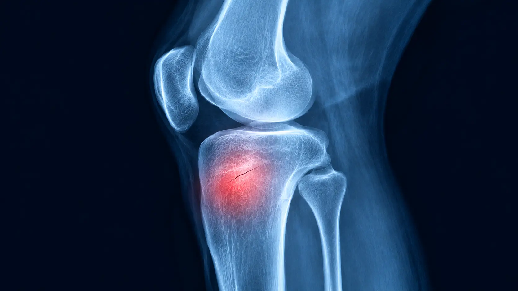
In active individuals, a knee stress fracture often develops when repeated loading puts more stress on healthy bone than it can repair. Common triggers include a sudden increase in running distance, speed or hill training, or changes in footwear or running surfaces. The upper shinbone (tibia), just below the knee, is a common site, particularly on the inner side. Because knee stress fracture symptoms can resemble those of a meniscal tear or tendonitis, the injury may initially be misdiagnosed as a problem affecting the meniscus or inner knee tendons.
The Person Over 55 With Sudden, Severe Knee Pain
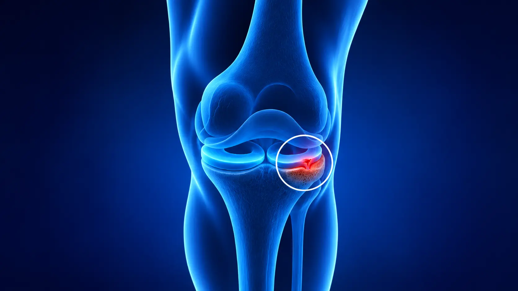
A subchondral insufficiency fracture of the knee is a different type of bone stress injury. It occurs in the bone just beneath the joint cartilage, where weakened bone may struggle to withstand normal everyday loads. It is more common in women and adults over 55. Pain may develop suddenly or worsen over several days, even without a fall or twisting injury. It typically gets worse when putting weight on the knee and may also disturb sleep. Older medical reports sometimes refer to this condition as spontaneous osteonecrosis of the knee (SONK).
This type of knee stress fracture can be mistaken for osteoarthritis or a meniscal tear, but the distinction matters because treatments suitable for these conditions, such as weight-bearing strengthening exercises, may not be appropriate. One study of middle-aged adults treated for suspected meniscal tears estimated that around 3 in 100 had an undiagnosed subchondral insufficiency fracture. Continuing to put excessive load on the knee may lead to collapse of the joint surface in some cases, making early diagnosis and appropriate treatment important.
The Rare Kneecap Stress Fracture
Stress fractures of the kneecap (patella) are rare and occur most often in athletes who engage in frequent running, jumping or kicking. Symptoms typically include gradually worsening pain at the front of the knee, which can initially be mistaken for patellar tendonitis. Because the kneecap is considered a high-risk site for a knee stress fracture, the crack may widen if left untreated. Imaging is important when anterior knee pain persists or does not follow the typical pattern, helping confirm the diagnosis and guide appropriate treatment.
What Causes Stress Fractures of the Knee?
Regardless of age, a knee stress fracture develops when the stress placed on the bone exceeds its ability to repair and rebuild itself.
Bone Stress Injury Knee Risk Factors
Several factors can increase the risk of a knee stress fracture. Sudden increases in training intensity, running on hard surfaces and changes in footwear can place additional stress on the bone. Low bone density, vitamin D deficiency, inadequate nutrition relative to activity levels, hormonal changes such as menopause, and long-term steroid use can weaken bone strength. Having a previous stress fracture also increases the risk of another injury. Knee mechanics play a role too. For example, a meniscal tear can increase pressure on the underlying bone, which may explain why these two conditions often occur together.
Knee Stress Fracture Symptoms
Knee stress fracture symptoms vary with the type, but these are the features to look for:
- Pain in one spot that is worse with weight-bearing, stairs or impact
- Pain that starts earlier in each walk or run, and takes longer to settle
- Tenderness when you press directly over the bone
- Mild swelling, and sometimes slight bruising
- Night pain or pain at rest as the injury progresses
- Sudden, severe pain without any injury, particularly after 55
Early Signs of a Knee Stress Fracture
Three key questions can help identify a possible knee stress fracture.
- First, where is the pain? Can you pinpoint the sore spot with one finger? Second, when does it occur?
- Does the pain start earlier with each activity, and does it settle within a few hours or persist?
- Third, what has changed recently?
- Have you started a new training programme, taken up a job that involves prolonged standing, or experienced a fall?
A bone stress injury often causes localised pain that progressively worsens and is closely linked to activity or weight-bearing. Recording your symptoms and recent changes before your appointment can help us assess the problem more efficiently.
Can You Walk With a Stress Fracture in Your Knee?
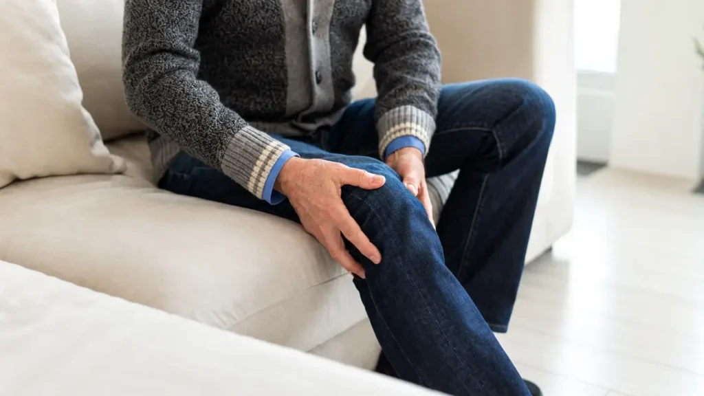
You may be able to, but pain is the signal to stop pushing. In the runner pattern, impact is the main trigger. In the over 55 pattern, even ordinary weight-bearing can hurt. Walking through the pain, or doing loaded exercises to strengthen the knee, can make a stress injury worse. If walking, stairs or squatting bring on a sharp or deep ache, ease off and get assessed rather than testing it repeatedly.
Why a Knee Stress Fracture Gets Mistaken for Something Else
A torn meniscus can mimic a stress fracture, and so can patellofemoral pain or a flare of knee osteoarthritis. They can also exist together. The table shows the clues that point away from one and towards another.
| Condition | Usual story | Clue to look for |
| Knee stress fracture | Load-related bone pain, sometimes night pain | One tender spot over the bone, normal early X-ray, worse with continued loading |
| Meniscal tear | Twist injury, catching or locking | Mechanical symptoms, though a tear can sit alongside a fracture |
| Patellofemoral pain | Front-of-knee ache with stairs, squats and long sitting | Usually improves gradually with exercise therapy |
| Osteoarthritis flare | Stiffness after rest that eases within minutes | A sudden, sharp worsening is not typical and needs checking |
| Tendon problem | Ache over a tendon with activity | Tender point sits over soft tissue and settles with load management |
Quick Comparison, Overuse Ache or Bone Injury?
| Question | Usually an overuse ache | Bone injury more likely |
| Does rest settle it within a day or two? | Yes | It lingers or returns sooner each time |
| Do several weeks of exercise or physio help? | Gradually | No change, or it gets worse |
| Where is it tender? | Spread over muscle or tendon | One spot over the bone |
| Does it wake you at night? | Rarely | Yes |
When Should You Seek Medical Advice for a Knee Stress Fracture?
Please get your knee assessed if several weeks of rest or physiotherapy have not helped, if exercise makes the pain worse, if the pain wakes you at night, or if an X-ray was normal but the pain carries on. Sudden severe pain with no injury, especially after 55, deserves a prompt appointment too. We can assess this quickly, explain what is happening and guide you on the next steps.
Symptoms That Need Prompt Assessment
Get in touch with us straight away if you have:
- Sudden severe knee pain
- Inability to put weight on the leg
- A visibly deformed knee or leg
- Rapidly worsening swelling
- Pain following a fall or direct injury
- Fever or feeling generally unwell with a hot, swollen knee
What Happens if a Knee Stress Fracture Is Left Untreated?
The risk depends on the pattern. In an athlete, a small crack that keeps being loaded can become a complete fracture and a much longer break from activity. In an insufficiency fracture, continued full loading can let the joint surface collapse, and end-stage damage may eventually need a knee replacement. Neither outcome is inevitable, and both become less likely when the injury is found early.
How Is a Knee Stress Fracture Diagnosed?
We start by listening. We ask how the pain behaves, what has changed in your activity, and about your bone health, medicines and any previous knee problems. We then examine the knee, checking where it is tender and how the joint moves and tracks. This tells us which scan, if any, will answer the question, and we help you understand what the results mean.
Why a Knee Stress Fracture Can Look Normal on an X-Ray
An X-ray shows changes that have already formed in the bone, such as new bone growth or a visible fracture line. In the early stages these have not appeared, so a stress fracture can be invisible for the first few weeks. A normal X-ray therefore does not rule one out. It is still useful, because it shows leg alignment and arthritis, and a repeat X-ray later can show healing.
Can an MRI Detect a Knee Stress Fracture?
Yes. MRI is highly sensitive for early bone stress injuries and is usually the preferred scan when an X-ray is normal but a stress fracture is suspected. It shows changes in the bone marrow before an X-ray can, and it also shows the meniscus, cartilage and ligaments, so other causes of pain can be checked at the same time. The right imaging pathway still depends on the injury we suspect.
Knee Stress Fracture MRI Findings Explained
MRI reports use technical words. This table translates the ones we see most often in a knee stress fracture MRI.
| What the report says | What it means in plain English | Why it matters |
| Bone marrow oedema (or bone marrow lesion) | Extra fluid-like signal in the bone, which looks bright on the scan | Shows the bone is stressed, but not the cause on its own |
| Bone bruise or contusion | Same appearance, linked to a recent knock or twist | A stress injury and a bruise can look alike, so your history matters |
| Stress reaction | Marrow oedema with no fracture line | An early injury that often needs a shorter recovery |
| Low-signal line or fracture line | A thin dark line through the bone | Confirms a true stress fracture |
| Cortical break | The crack has gone through the outer wall of the bone | A more advanced fracture that needs closer protection |
| Subchondral insufficiency fracture | A fracture line just under the cartilage, in weakened bone | The over 55 pattern, once called SONK |
| Subchondral collapse or flattening | The bone under the joint surface has given way | Needs prompt specialist review |
| Meniscal tear also noted | A tear sits alongside the bone injury | We work out which is causing most of your pain |
No single phrase makes the diagnosis. The scan is always read alongside your story and examination.
When a CT Scan or Bone Scan Is Used Instead
A CT scan shows the fine detail of a fracture line when the picture is unclear. A bone scan is used less often, usually if MRI is not possible or if a stress fracture is still suspected after an MRI that shows nothing.
Knee Stress Fracture Treatment
Knee stress fracture treatment follows the location and type of the injury, and the same words, such as rest and protected weight-bearing, mean different things in different places. This is a general guide, and your own plan will depend on your scan, your examination and what you want to return to.
| Where the bone is injured | Who it usually affects | How it is usually approached |
| Upper shin, including the tibial plateau | Runners and other athletes | Reduce load, with crutches or a brace for a short time in some people, then stepwise loading |
| Lower thighbone, under the cartilage | Mostly over 55, sometimes athletes | Protected weight-bearing for weeks, close follow-up and a bone health review |
| Kneecap | Rare, jumping and running athletes | Specialist-led, as a higher-risk site. Some heal without surgery, others need fixation |
| Joint surface that has collapsed | Delayed or missed insufficiency fractures | Surgical options are discussed, from joint preservation to replacement |
Activity Modification and Load Management
For most people, treatment starts by stopping the activities that cause pain and swapping in lower-impact options your specialist approves. Pain is the guide, and activity only goes up once daily walking is comfortable. Simple pain relief may be suggested, but it should not be used to push through training.
Do You Need Crutches, a Brace or Immobilisation?
Not everyone does. Some people only need to reduce their activity. Others benefit from crutches or a stick to offload the knee, and a smaller group needs a brace or a short period of immobilisation. For insufficiency fractures there is no single agreed schedule, and published approaches range from several weeks to a few months of protected loading. We explain the plan that suits your fracture and review it as you improve.
When Does a Knee Stress Fracture Need Surgery?
Most knee stress fractures heal without an operation. Surgery is considered when a fracture is displaced or unstable, sits in a higher-risk site, fails to heal despite proper treatment, or when the joint surface has collapsed. It is not automatic for every complete fracture. In selected people whose leg alignment keeps overloading one side of the knee, Mr Raghbir Khakha, who takes a joint preservation approach, may also discuss whether a knee osteotomy has a role in a joint-preserving plan, and we explain the benefits and limits of every option.
Physiotherapy, Biomechanics, Nutrition and Bone Health
Healing the bone is only half the job. Supervised physiotherapy rebuilds strength and control once the bone allows it, and a review of your running technique, footwear and training pattern helps prevent a repeat. Eating enough for your activity level, with adequate calcium and vitamin D, supports repair. If weaker bone is suspected, we may suggest blood tests or a bone density scan, and we discuss whether any bone-strengthening treatment is appropriate. Please check with us before starting high-dose supplements.
Knee Stress Fracture Recovery Time
How Long Does a Knee Stress Fracture Take to Heal?
The honest answer is weeks for some people and months for others. Many low-risk fractures in athletes settle in roughly 6 to 12 weeks, though some take longer. Insufficiency fractures heal more slowly, often over several months. These are general guides, not promises, and your own timeline depends on the location, the severity and how you respond.
What Affects Healing Time?
The main factors are:
- The type and location of the fracture
- How early it was diagnosed
- How well load is managed during recovery
- Bone health, vitamin D and nutrition
- Age, hormonal factors, smoking and body weight
- Whether you have had stress fractures before
Signs Your Knee Is Healing
The signs are pain-free walking, tenderness over the bone fading, and nights without aching. Some people have a follow-up MRI that shows the changes resolving, though not everyone needs one. We decide on follow-up imaging case by case.
How Do You Return to Walking, Exercise and Sport After a Knee Stress Fracture?
The return depends on your pattern. Runners usually progress from pain-free walking to low-impact conditioning, then walk-run intervals, changing only one thing at a time, such as distance or speed. After an insufficiency fracture, the early focus is low-impact activity, and loaded exercises such as deep squats wait until you are cleared. Anyone returning from knee running injuries or other knee sports injuries and trauma should expect a gradual build up. If pain returns at any stage, step back and let us know.
Book a Knee Stress Fracture Assessment at London Knee Care
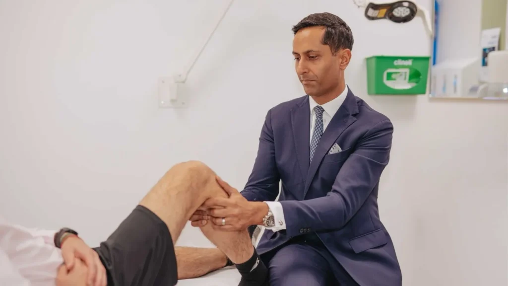
Mr Raghbir Khakha is a Consultant Orthopaedic and Specialist Knee Surgeon at London Knee Care in London. He has been in consultant practice since 2017 and takes a joint preservation approach to knee care. His areas of focus include meniscal tears, sports injuries such as ACL and PCL injuries, and young knee preservation.
If your knee has not responded to the usual treatment, a normal X-ray should not be the end of the conversation. We start with a clear diagnosis, so your treatment is based on what is happening in the bone rather than a standard protocol. We assess your symptoms, arrange the right imaging, explain your MRI report in plain language and guide your protected loading and rehabilitation.
Our new Acute Knee Clinic also offers prompt assessment of acute knee injuries on Saturdays at the Cromwell Hospital from November. For details and availability, contact us and we will help you find the right time. If the knee suddenly becomes severely painful, you cannot put weight on the leg, or it looks deformed, get in touch with us the same day.
Address: 45 Queen Anne Street, London W1G 9JF
Phone: 0207 046 8008
Email: rk@os.clinic
Frequently Asked Questions About Knee Stress Fracture
What Are the Early Signs of a Knee Stress Fracture?
Pain in one spot that starts earlier each time you walk or run, tenderness when you press over the bone, and pain that does not settle with rest are the usual early signs. Mild swelling can appear too. Night pain is a later sign, so do not wait for it before getting the knee assessed.
Can a Knee Stress Fracture Be Missed on an X-Ray?
Yes. Early stress fractures often do not show on an X-ray, because the bone changes have not formed yet. That is why an MRI is commonly used when suspicion stays high. A normal X-ray does not rule a stress fracture out, and a repeat X-ray later can show healing.
Can a Knee Stress Fracture Feel Like a Torn Meniscus?
Yes, and the two can occur together. Catching or locking points more towards a meniscal tear, while a tender spot over the bone and worsening pain with load point towards a fracture. An MRI helps tell them apart.
Does a Knee Stress Fracture Require Crutches?
Not always. Some people need crutches, a stick or a brace to offload the knee, while others only need to change their activity. It depends on the fracture site and whether the fracture is stable, which is why the plan is set after your scan and examination.
When Can You Return to Exercise After a Knee Stress Fracture?
You can usually return once walking is pain-free, the bone is no longer tender and we agree it is safe. The return is gradual, with one change at a time, and the timing varies from person to person depending on the type and location of the fracture.
This article is general information and does not replace a personal assessment. If you are worried about your knee, please get in touch with us and we will arrange your assessment.